Wolfner Lab
Department of Molecular Biology and Genetics
Cornell University
Research
Broadly, our lab is interested in using molecular biology and genetics to dissect the important reproductive processes that occur around the time when a sperm fertilizes an egg. We use the fruit fly, Drosophila melanogaster, for most of our work. Drosophila reproduction and development can be readily studied with molecular genetic and genomic techniques. Furthermore, Drosophila serves as a model for other animal systems. Many of the genes and reproductive/developmental phenomena in flies have counterparts or analogues in other animals, including humans and insect vectors of disease.
Seminal fluid proteins
Seminal fluid proteins (Sfps) are important for fertility in all animals, including people. They play particularly striking roles in insects, where they affect the behavior and physiology of the female. Studying Sfps' functions provides insights into the molecular and genetic mechanisms of reproduction. Sfps are also relevant evolutionarily, as they impact sperm competition and often evolve rapidly and adaptively between species. Work in insect Sfps can provide information to help design ways to control the reproduction of harmful insects, such as vectors of disease.
Figure: Sperm mass stained for a seminal fluid protein (green) and nuclei (blue). Some sperm also express ProtamineB-RFP (red). See Misra and Wolfner 2020.
%20and%20nuclei%20(blue)_.jpg)
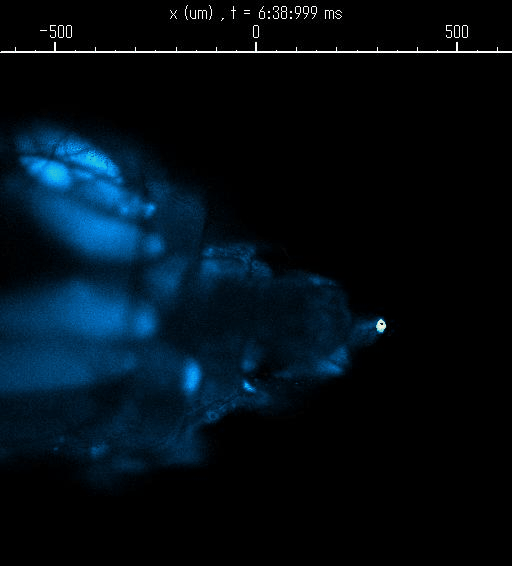
Egg Activation
Mature oocytes are arrested at meiosis before the start of embryo development, waiting to be awakened to fulfill its roles as totipotent zygotes. This "activation" process is highly conserved across species, and is universally accompanied by a rise in intracellular Calcium (Ca2+). In Wolfner Lab, we use Drosophila as a model organism to study the detailed mechanism underlying the Ca2+ wave during egg activation and how intra- and extracellular cues might regulate this process.
Figure: An oocyte that expresses a calcium sensor undergoing activation when passing through the oviduct. See joint article with the Aigaki lab, Kaneuchi et al. 2015.